Dental X-rays are a proven diagnostic tool that helps dentists detect oral health issues, such as cavities, fractures, or gum disease. There are both intraoral X-rays which are those taken inside the mouth, and extraoral, which are taken outside the mouth. With X-rays, dentists can examine tooth structures that they aren’t able to see during a routine examination, such as the jawbone, nerves, gum tissue, sinuses, and teeth roots.
In a quest to deliver higher quality and safer dental care, many dentists have embraced the transition from traditional film X-rays to digital X-rays. These offer advanced imaging techniques that eliminate some of the shortcomings of traditional film and are also a safer alternative. In this article, we will explore what makes digital X-rays superior, from image quality to less environmental waste.
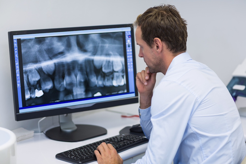
Image Quality
In order for a dentist to make a proper diagnosis and determine the most effective treatment plan, they need clear images of the dental issue. Digital X-rays offer a higher quality image than traditional film X-rays, to the point of allowing a dentist to see the tiniest of fractures and irregularities that are often missed with traditional film. Digital radiography machines are equipped with software that greatly improves image definition, in addition to allowing the dentist to zoom in or adjust the contrast for a better look. They offer more than 200 shades of gray, while traditional X-rays have only 16-25 shades of gray. The enhanced visuals of digital X-rays can help dentists identify oral health issues earlier, possibly saving the patient time, money, and discomfort.
Processing Time
Processing time has always been a negative aspect of traditional X-rays, considering the wait time for the film to develop before images could be viewed. Conversely, digital images are available for viewing instantly. This allows a dentist to evaluate the situation immediately and provide a diagnosis during that same appointment. The quicker turnaround time reduces the number of visits needed and allows for a faster start to whatever treatment is needed. The short timeline in viewing images also allows dentists to see more patients in a single day, reducing the wait time to see a dentist when a patient is experiencing an issue.
Reduced Resources
Traditional X-rays require more resources than digital X-rays, with chemical developers and film processors needed as part of the process. Digital X-rays are just a one-time investment in equipment, with dental hygienists and assistants the only staffers needed to take X-rays and confirm the quality on a computer. This means less labor in a dental office and also extra space since there is no need for green rooms or radiology rooms.
Ease-Of-Use
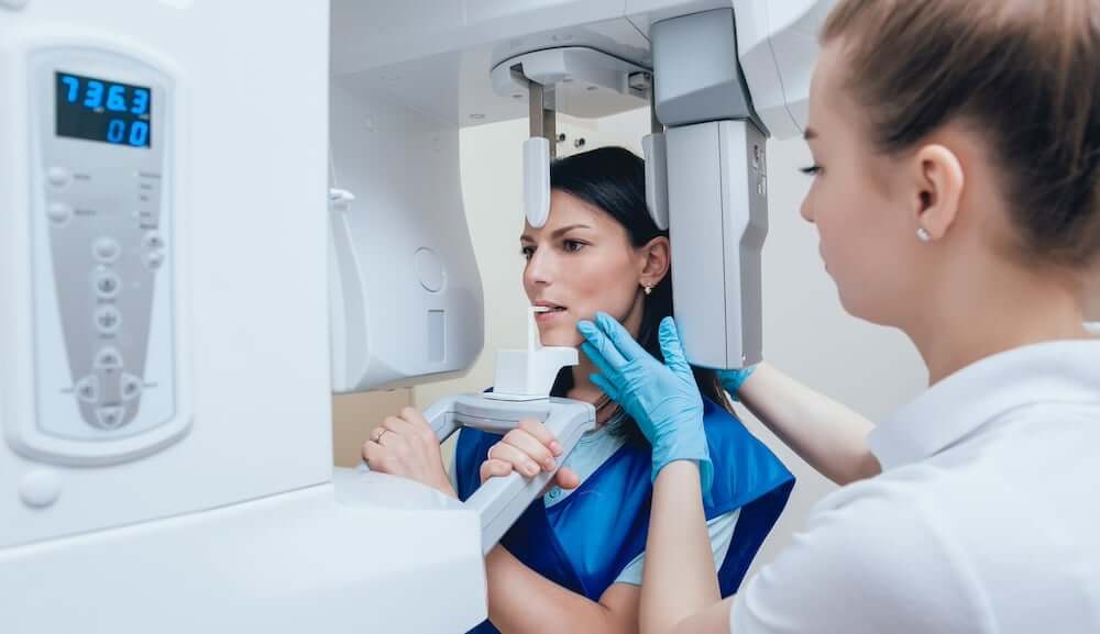
Digital X-rays require only minimal training, with operators trained quickly and with full confidence in operating the machines. They are very straightforward compared to traditional radiography, with dentists agreeing that it is also a cleaner and faster process. The digital process involves a lead apron being placed over the patient’s chest, the mouthpiece positioned according to the tooth or teeth requiring imaging, and then the dentist, hygienist, or dental assistant simply presses a button to capture the image. The image immediately appears on a computer screen so the clarity and appropriate positioning can be confirmed.
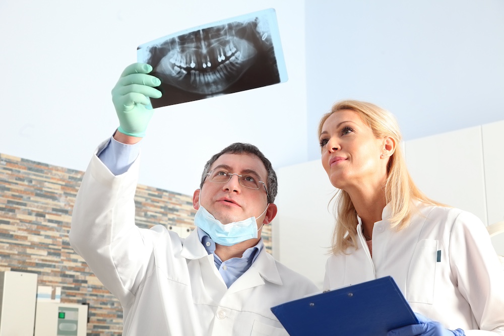
Image Sharing & Storage
Two of the most significant differences between traditional and digital dental X-rays is how the images are shared and stored. Traditional X-rays are synonymous with carrying around large envelopes containing multiple films. If a dentist needs to consult with another medical professional or if a patient is switching dentists, those bulky folders need to come along. With digital X-rays, dentists can electronically share the images with other experts minus the folder of films, and without any image quality loss. Likewise, with a patient transferring to a new dentist, sharing their X-ray history can be done electronically.
Digital X-rays are also much easier to store than films, with far less chance of them being lost, misplaced, or misfiled. The images are stored on a protected hard drive with limited access, and retrieving them is as easy as pulling up a computer file.
Patient Safety
A common concern among dental patients is the radiation exposure that comes with X-rays. According to The Cleveland Clinic, digital X-rays use 80-90% less radiation, making it incredibly low exposure compared to traditional X-rays. Radiation exposure to digital dental X-rays is similar to the minimal exposure from televisions, smartphones, and computers. In addition, due to the detail and accuracy offered by digital images, there is minimal chance of needing to retake a dental X-ray and therefore being exposed to more radiation. Retakes are a fairly common occurrence with traditional film X-rays, as the clarity and proper positioning are often unknown until the films are developed.
Environmental Waste
In today’s environmentally-conscious world, another benefit of digital versus traditional film X-rays is the type and volume of waste produced. X-ray film is made of PET plastic and is known to contain toxic chemicals that can leach into waterways after being placed in landfills. These materials can lead to damage to the skin, lungs, and eyes. Also, a by-product of X-ray film is lead foils, which can stay in topsoil for 2,000 years and, therefore, easily infiltrate our food ecosystem. According to the Eco Dentistry Association, these toxins remain an environmental threat, as only 60% of U.S. dental offices have eliminated these materials from their practices by switching to digital imaging. With the absence of physical film when using digital X-rays, there is no concern about film disposal and environmental consequences.
Contact us if you would like to learn more about digital versus traditional film X-rays.