Dental X-rays are the go-to diagnostic tool used by dentists to detect cavities, fractures, or gum disease. With X-rays, dentists can examine tooth structures that they aren’t able to see during a visual examination, such as the jawbone, nerves, gum tissue, sinuses, and teeth roots. Today, most U.S. dentists have transitioned from traditional film X-rays to digital X-rays. Digital X-rays offer advanced imaging that has eliminated many of the shortcomings of traditional film, such as image quality, and is also considered a safer alternative.
Why Do I Need Dental X-rays?

Your dentist will take dental X-rays every year in order to get a clear picture of your overall oral health. In addition, they may be taken to help make a diagnosis if you’re experiencing a dental issue and to help determine the most effective treatment plan. Dental X-rays offer a high-quality image that allows your dentist to see even the smallest of tooth fractures and irregularities. X-ray machines are equipped with software that provides outstanding image definition and allows your dentist to zoom in or adjust contrast for an even better look.
Types of Dental X-Rays
There are both intraoral X-rays, which are those taken inside the mouth, and extraoral, which are taken outside the mouth. Both intraoral and extraoral X-rays include various types.
The different types of intraoral X-rays are:
-
Bitewing
-
Periapical
-
Occlusal
The different types of extraoral X-rays are:
-
Panoramic
-
Cephalometric
-
Cone beam
Intraoral X-Rays
Bitewing X-rays help your dentist detect decay between your teeth and just below the gum line. They show the upper and lower teeth in one specific area of your mouth but do not typically show the roots of your teeth.
Periapical X-rays help your dentist detect decay, gum disease, bone loss, and other significant abnormalities in the teeth and surrounding bone. They show the entire tooth, from the crown down to the root tip.
Occlusal X-rays help your dentist detect issues in the floor and roof of your mouth, and help to diagnose fractured teeth, impacted teeth, and problems with the front teeth roots. They can also detect cysts, abscesses, and jaw fractures.
Extraoral X-Rays
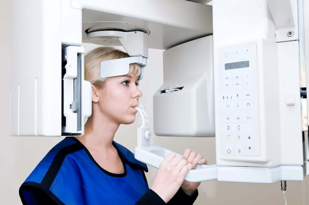
Panoramic X-rays provide your dentist with an overarching look at your dental health. They show all structures in your mouth in a single, wide image, including the upper and lower teeth, jaw joints, nerves, sinuses, and bone.
Cephalometric X-rays help a dentist or orthodontist plan treatment for correcting bites. They show the entire head from a side angle, demonstrating the position of teeth in relation to the jaw.
Cone beam is a type of CT scan that dental surgeons use to confirm the height, width, and location of the jawbone before a dental implant procedure. It captures 3D dental X-rays of the teeth, jaws, joints, nerves, and sinuses and can also detect tumors and facial fractures.
If you have additional questions about dental X-rays and the various types used, contact us.