Modern dentistry is synonymous with advanced technology, which has significantly enhanced diagnostic capabilities and treatment planning for patients. Among the innovations that have redefined dentistry is dental cone beam computed tomography (CT) scans — a revolutionary tool that provides detailed 3D images of the oral and maxillofacial region. From pinpointing complex dental issues to aiding in surgical planning, cone beam CT scans have become commonplace in dental practices.
What is a Dental Cone Beam CT Scan?
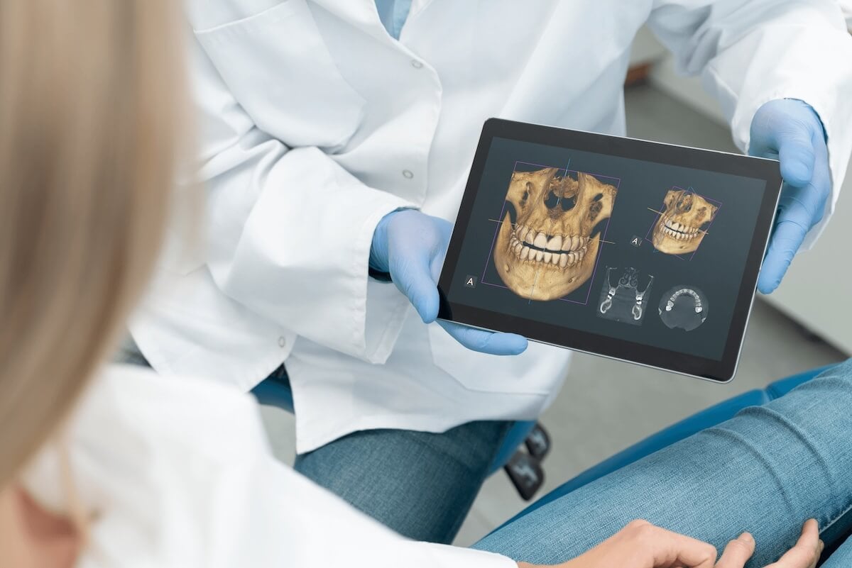
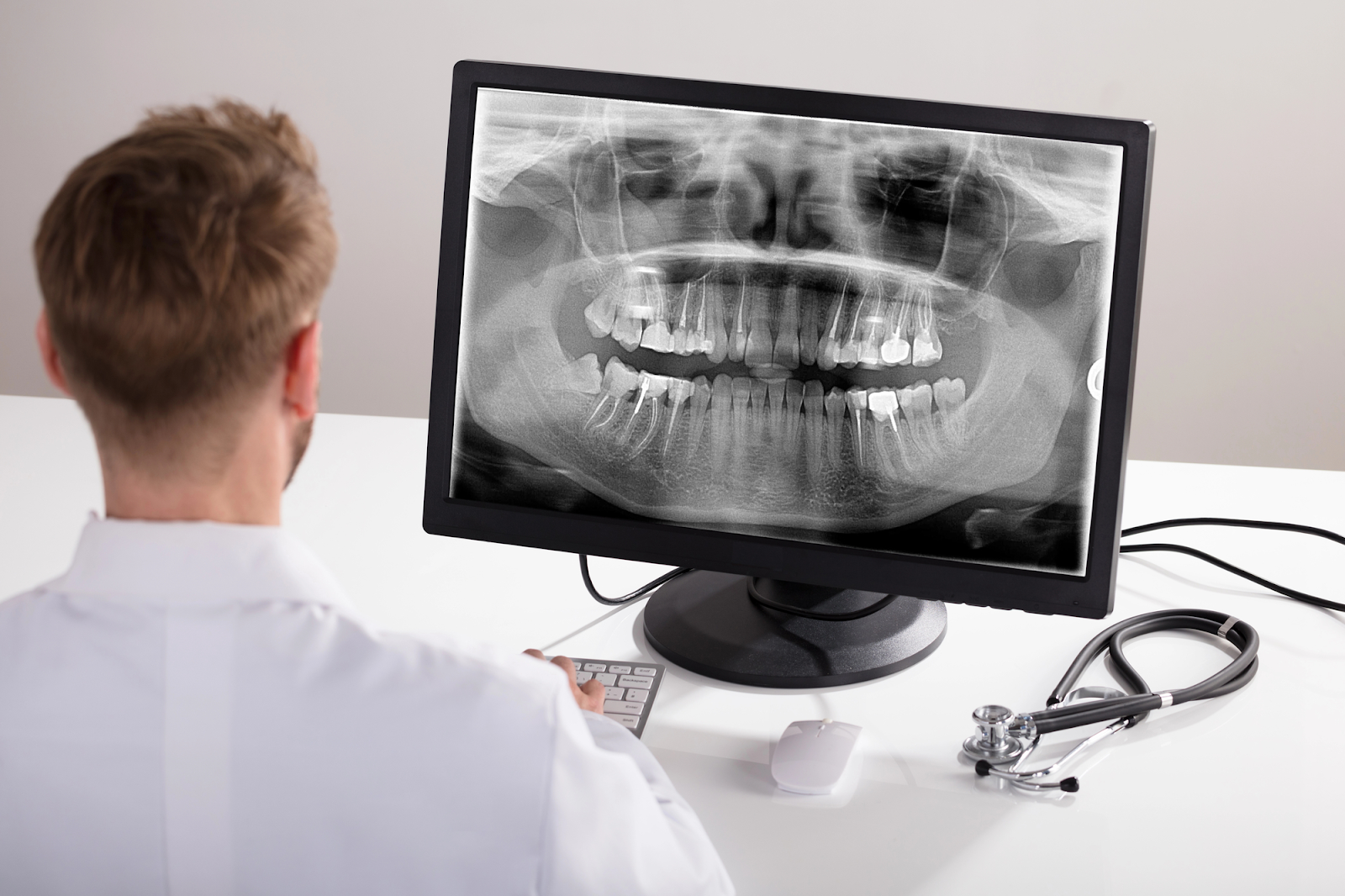
A dental cone beam CT scan (CBCT) is a specialized imaging technique that uses cone-shaped X-ray beams to create a 3D representation of oral structures. Unlike traditional dental X-rays, which produce 2D images, cone beam CT scans provide very detailed, high-resolution images with minimal distortion. This technology allows dentists to view the teeth, jawbone, nerves, sinuses, and surrounding tissues from various angles, offering a more comprehensive view of the patient's oral anatomy and any issues.
When is a Dental Cone Beam CT Scan Necessary?
The need for a dental cone beam CT scan varies depending on the patient's specific dental needs. While some patients may require periodic imaging for ongoing treatment monitoring, others may undergo CBCT scans on a one-time basis for a particular diagnosis or treatment planning.
Dental cone beam CT scans are utilized in a variety of scenarios, including:
-
Implant Planning: Before dental implant placement, cone beam CT scans help assess bone quality, quantity, and location, aiding in implant positioning.
-
Orthodontic Treatment: Orthodontists utilize CBCT images to evaluate impacted teeth and plan orthodontic interventions.
-
Evaluation of Pathologies: CBCT scans are critical in diagnosing and evaluating cysts, tumors, infections, and temporomandibular joint (TMJ) disorders.
-
Surgical Planning: Oral surgeons rely on cone beam CT scans for accurate pre-surgical planning, for procedures such as impacted wisdom tooth removal and bone grafting.
How Does a Cone Beam CT Scan Work?
A cone beam CT scan is straightforward and typically only takes a few minutes. The patient is positioned standing or seated within the CBCT machine, which resembles a traditional dental X-ray unit, but with a rotating arm. The machine emits a cone-shaped X-ray beam that rotates around the patient's head, capturing multiple images from different angles. These images are reconstructed into a 3D digital model using specialized software, which can then be viewed and manipulated by the dentist or radiologist.
What Diagnoses are Made From Cone Beam CT Scans?
Cone beam CT scans help dentists diagnose several oral health issues, along with a visual examination and understanding of the patient’s symptoms.
Dental professionals gather considerable information from cone beam CT scans, including:
-
Bone density and structure issues
-
Problematic eruption patterns of teeth
-
Soft tissue structure issues
-
Cysts, tumors, and abscesses
-
Impacted teeth
-
Oral infections
Are Cone Beam CT Scans Safe for Children?
While dental cone beam CT scans are commonly performed on adults, they are also valuable diagnostic tools in pediatric dentistry. Children may undergo CBCT scans for various reasons, including assessing dental and skeletal development, evaluating impacted teeth, diagnosing craniofacial anomalies, and planning orthodontic treatment. However, due to concerns regarding radiation exposure, pediatric dental professionals carefully weigh the benefits and risks of CBCT imaging in children.
Contact us to learn more about dental cone beam CT scans, a form of imaging that has become an indispensable tool in modern dentistry.